I’ll again be looking at some of the myths surrounding back pain and our spine. If you haven’t already, I advise you to read part 1 as in it I explain why these myths can be bad for you and why I think it is important to address them. In it, I have given an explanation on serious back pathologies being rare and that our back hurting often does not mean we have injured something.
Today I want to look at MRI scans, x-rays and the sort of investigations that usually give us an actual picture of our back, and try to understand what they role is in the management of back pain.
It may have been that some of you have been to your doctor or physiotherapist due to having really bad pain on your back. It was so bad that it felt like something inside your spine came out or was crushed – something bad happened surely. But when you went to your doctor they didn’t mention having a scan or even told you they don’t need one. How can they now what is wrong if they don’t use a scan to look into the actual spine?
The problem with this line of reasoning is that scans can’t show us pain.
Scans can show us the appearance of structures – bones, ligaments, cartilage, fluid, fat – in our body. Some of these structures are expected to appear a certain way in a scan, be it in terms of shape, size, how dark or bright they look. The problem starts when we look at the data investigating what looks “normal” (a word I would argue that has very limited value in healthcare) and what doesn’t.
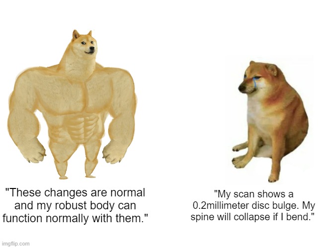
A study by Brinjikji et al (2015) looked at how common some so called structural changes – changes in how the brightness of a disc looks in a scan, the size of the discs in our spine, changes in the shape of the joints in our spine – in people without any pain. They found that even in people who are 20 years of age, disc bulges were present in 30% , disc protrusions in 29% and disc degeneration in 37%. These values tend to increase with age, with disc bulges and degeneration being present in over 70%, reduction of disc thickness in 56%, degeneration of joint facet in 32% and disc fissures in 23% of people who are 50 years old.
In another review by Teraguchi et al (2018), the discs with brighter than normal zones on MRI were found in between 20-24% of people. In a more recent population-based study by Kasch et al (2020) limited to Germany but with a large sample 3369 people followed from 2008 to 2019, 74.4% of people without pain had at least one finding on MRI.
Even something as scary as spondylolisthesis, actual sliding of a vertebral body in relation to the ones around it as a result of a small fracture (and no, this is not what people mean when your vertebrae come out of alignment nor it can or should be treated with manipulation or manual therapy of any kind) will be present in 50% percent of people who are 80 years of age (Brinjikji et al, 2015). And in case you had forgotten, this is all in people without any symptoms – no pain, no leg tingling, nothing.
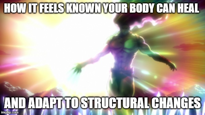
The next thing to consider with scans to our spine is that some of the changes that can be seen also often return to normal if we just wait.
In a systematic review by Chiu et al (2015) it was found that herniated discs will reduce over time, from 13% of minor bulges reducing and 11% completely resolving, to 96% of the very serious disc herniations where part of the disc has almost fully separated from the main disc reducing in size and 43% completely resolving. You read that right: the worse ones are much more likely to improve without any sort of treatment.
The last thing to consider is if having a scan would change how we treat back pain. As we have seen so far, what the scan shows and what symptoms you get or how bad they are don’t really match up. Looking again at the study by Kasch et al (2020), after following up people and comparing their symptoms at baseline with how they presented after 11 years, the MRI findings did not match or predict the severity or presence of symptoms. Based on this information and referring specifically to back pain, because we are not able to match what is shown on a scan with your pain, we also can’t really use it to inform how we are going to treat you. A systematic review by Karel et al (2015) showed that having an early scan did not offer any improvement on pain, function, satisfaction, quality of life and overall improvement in people with back pain, on either short- or long-term follow up. As argued by Brinjikji et al (2015), this all suggests that these findings are just part of life and the natural processes of our body rather than something pathologic that needs treatment. Isn’t our body amazing and resilient?
I hope that what I have described so far has helped you understand why when being assessed by a health professional, unless certain other symptoms are present besides pain, they will try discouraging you from having a scan to your back. At least they will try to do this if they are up to date with the major present clinical guidelines, which all advise against routine use of scans for back pain (Cuff, 2020). And believe me in that if there are any symptoms of anything more serious being present – which pain on its own isn’t – your doctor or physiotherapist will suggest it before you.
We – both clinicians and members of the public – often think that scans can be this miraculous window into our body that will tell us straight away what is happening. However, as we have established, pain is a complex and non-straightforward thing. Even very high-tech scans are surrounded by a lot of uncertainty when it comes to showing causes of pain.
I hope this text has helped you understand a bit more about where this uncertainty comes from and what the research shows on the effectiveness and usefulness of scans. If you are a member of the general public and the clinician you’re seeing advises against a scan, take is as an honest understanding of how little it may help. If you are a healthcare professional, use the research cited to better understand the limitations of the imaging technology we have available and to avoid over-medicalizing what are actually normal findings in healthy people. We have a responsibility to be well informed and not give bad advice to the people who come to us for help.
As always, share this text throughout social media if you felt you learned something from it. Feel free to ask any questions in the comments and I hope you will return for the next one.
References:
Brinjikji, W., Luetmer, P. H., Comstock, B., Bresnahan, B. W., Chen, L. E., Deyo, R. A., Halabi, S., Turner, J. A., Avins, A. L., James, K., Wald, J. T., Kallmes, D. F. and Jarvik, J. G. 2015. Systematic Literature Review of Imaging Features of Spinal Degeneration in Asymptomatic Populations. Am J Neuroradiol, 36(4), pp.811-16 DOI: https://doi.org/10.3174/ajnr.A4173
Chiu, C.-C., Chuang, T.-Y., Chang, K.-H., Wu, C.-H., Lin, P.-W., & Hsu, W.-Y. 201. The probability of spontaneous regression of lumbar herniated disc: a systematic review. Clinical Rehabilitation, 29(2), 184–195. doi:10.1177/0269215514540919
Cuff, A., Parton, S., Tyer, R., Dikomitis, L., Foster, N. and Littlewood, C. 2020. Guidelines for the use of diagnostic imaging in musculoskeletal pain conditions affecting the lower back, knee and shoulder: A scoping review, Musculoskeletal Care, 18(4), pp. 546–554. doi: 10.1002/msc.1497.
Jensen, R. K., Jensen, T. S., Koes, B. and Hartvigsen, J. 2020. Prevalence of lumbar spinal stenosis in general and clinical populations: a systematic review and meta-analysis, European Spine Journal. Springer Berlin Heidelberg, 29(9), pp. 2143–2163. doi: 10.1007/s00586-020-06339-1.
Kasch, R., Truthmann, J., Hancock, M. J., Maher, C. G., Otto, M., Nell, C., Reichwein, N., Bülow, R., Chenoti, J.-F., Hofer, A., Wassilew, G. and Schmidt, C. O. 2021. Association of Lumbar MRI Findings with Current and Future Back Pain in a Population-Based Cohort Study, Spine, 0. doi: 10.1097/BRS.0000000000004198.
Teraguchi, M., Yim, R., Cheung, J. P.-Y. and Samartzis, D. 2018. The association of high-intensity zones on MRI and low back pain: a systematic review, Scoliosis and Spinal Disorders. Scoliosis and Spinal Disorders, 13(1), pp. 14–19. doi: 10.1186/s13013-018-0168-9.
